
1 What draws me to this area
I am interested in how we move and the tissues that are involved in movement. Walking and balance bring this connection into focus: whole-body movement depends on the interaction of joints, muscles, soft tissues and the ground beneath our feet.
My research experience spans clinical human-motion analysis at Cambridge and foot and ankle biomechanics at Liverpool. Together, these experiences have shaped my interest in studying movement at different scales, from head and whole-body motion capture to the structure and mechanical behaviour of individual tissues. I bring sensing, biomedical imaging and computational modelling together to explore those connections.
2 Questions that interest me
Some questions that motivate my work include:
- What can changes in walking and balance tell us, and how can we measure them reliably outside the laboratory?
- How do tissue structure, ageing and loading interact with the mechanics of movement?
- How can imaging and personalised models help us interpret differences between people?
These questions offer starting points for collaboration across engineering, biomechanics and clinically motivated research.
3 Understanding movement in a clinical context
At the University of Cambridge, I worked on head and whole-body movement analysis with medical consultants and clinical scientists at Addenbrooke’s Hospital (Cambridge University Hospitals NHS Foundation Trust). I contributed to the development of data processing pipelines for ear-based wearable devices for monitoring walking and activities of daily living, and planned optical motion-capture setups that are clinically relevant in the context of balance and neurological disorders.
A recurring question for me is what a measurement actually tells us: which features of movement are informative, how reliably they can be obtained, and how we can interpret them in relation to the person being assessed.
4 Looking beneath the surface
Ultrasound (US), bi-planar X-ray and magnetic resonance imaging (MRI) are important parts of my experience in biomechanics research. They offer complementary views of tissue structure, deformation and bone motion, helping connect external measurements with what happens within the body.
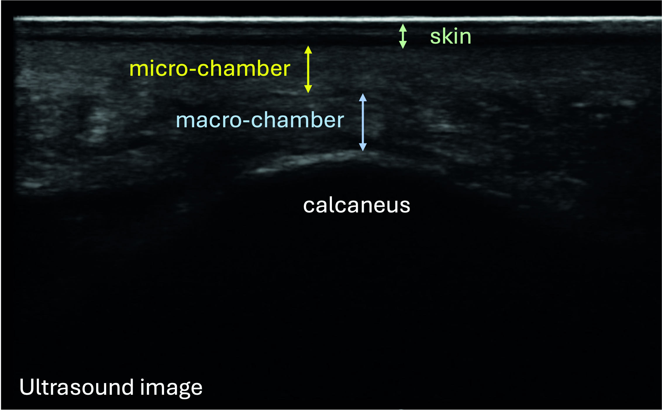
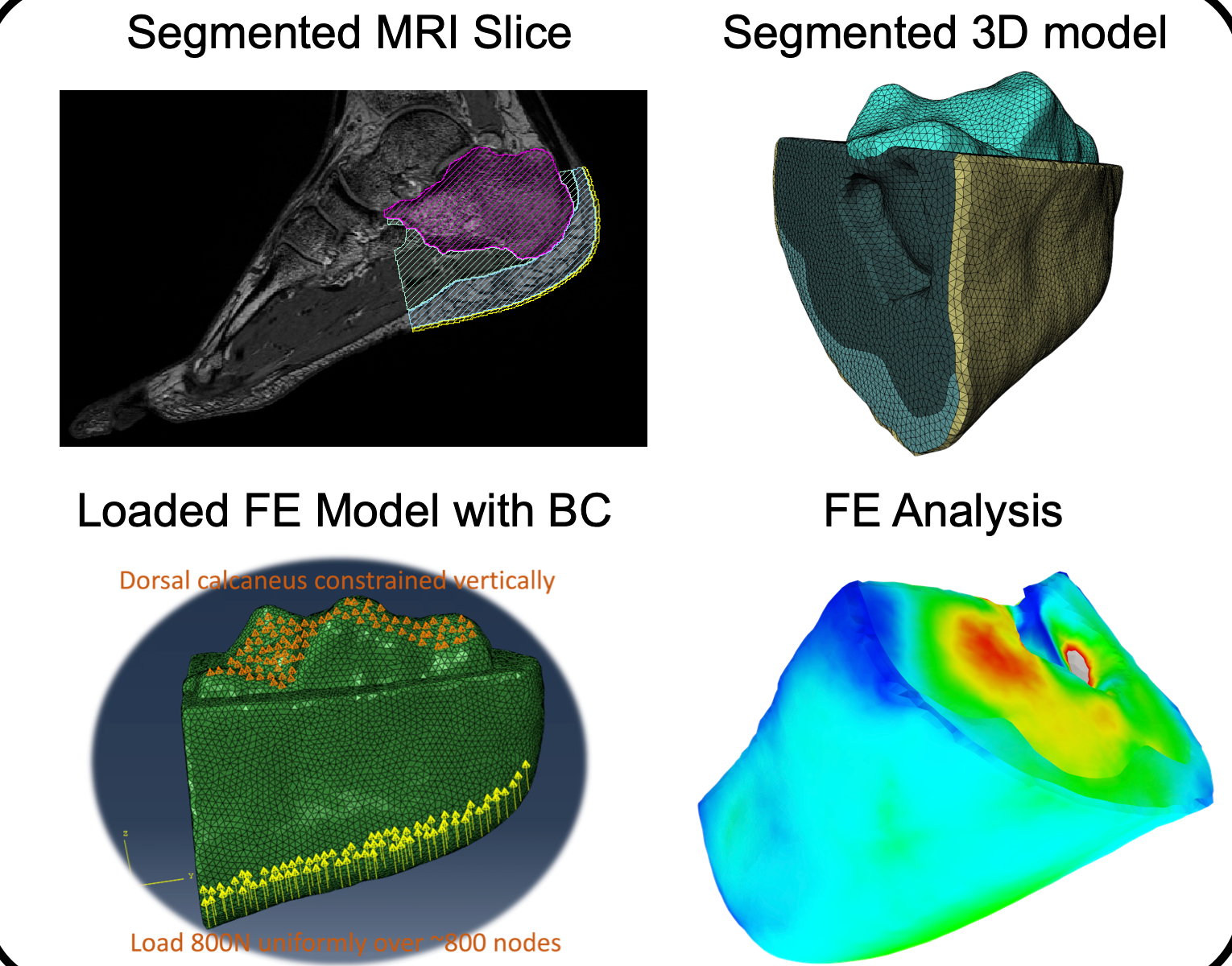
At Liverpool, I developed equipment for heel-pad ultrasound imaging and plantar-surface strain measurements. I also constructed participant-specific finite-element models from segmented foot MRI scans and conducted bi-planar X-ray videography experiments with simultaneous force-plate measurements to inform hindfoot simulations.
What draws me to this work is the possibility of linking anatomy with function. Ultrasound can reveal how the layers beneath the heel respond to loading; MRI provides anatomical information for personalised models; and bi-planar X-ray enables simultaneous investigation of motion and tissue behaviour. These approaches help explore mechanisms that are difficult to observe from the outside alone.
5 Contributions from my work
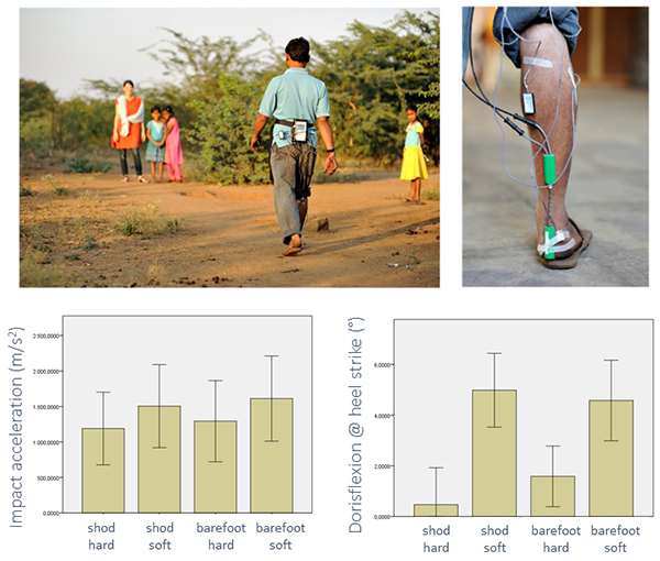
Working with colleagues at Liverpool, I contributed to participant studies in the UK, India and Namibia, combining tissue measurements, gait biomechanics and biomedical imaging. Our work included examining age-related differences in the layers of the heel pad. I also investigated spatiotemporal gait parameters across the three populations using both laboratory and field gait measurements, and their correlations with age, weight and other confounding variables.
I also explored a complementary question: can a quick measurement of surface hardness represent how heel tissues compress under load? Our conference work found that it did not reliably do so under the tested conditions. This illustrates a question I find especially important: when does a convenient measurement capture the behaviour we want to understand?
Alongside experimental work, I contributed to a critical review of footwear and locomotor health across the life course, examining the evidence behind common footwear features and their interaction with the foot’s own mechanics.
6 Connecting measurements and models
My approach combines wearable and optical sensing, force and pressure measurements, biomedical imaging, and musculoskeletal and finite-element modelling. I value their complementary perspectives: measurements describe movement and loading, imaging reveals anatomical structure, and models help investigate mechanical behaviour.
Across these activities, I aim to keep the analysis grounded in experimental evidence and attentive to variation between people.
7 Possibilities and connections
I would like to explore further connections between tissue biomechanics, whole-body movement and practical mobility assessment. Possible directions include combining wearable measurements with personalised models, studying variation across populations, and developing accessible ways to interpret biomechanical data.
I welcome conversations with researchers, clinicians and prospective research students interested in human movement, tissue mechanics, biomedical imaging or computational modelling. If your interests connect with these themes or bring a different perspective, I would be happy to hear from you.
.
8 Selected work
- Ageing and heel tissues: Impact of ageing on the heel pad: ultrasound imaging of the macrolayer, microlayer and skin — conference abstract, Footwear Science, 2025.
- Interpreting tissue measurements: Reliability of Shore Hardness as a Surrogate for Heel Pad Stiffness: A Correlative Study Using Ultrasound Imaging — ISB conference poster, 2025.
- Footwear and walking: Footwear Choice and Locomotor Health Throughout the Life Course: A Critical Review — Healthcare, 2025.
Figure credits
- Featured fieldwork figure: Evolutionary Morphology and Biomechanics Group, University of Liverpool, Figure 5.
- Heel-pad ultrasound: Elnaggar, Rowlatt and D’Août, ISB 2025 poster.